Pelvic Kidney
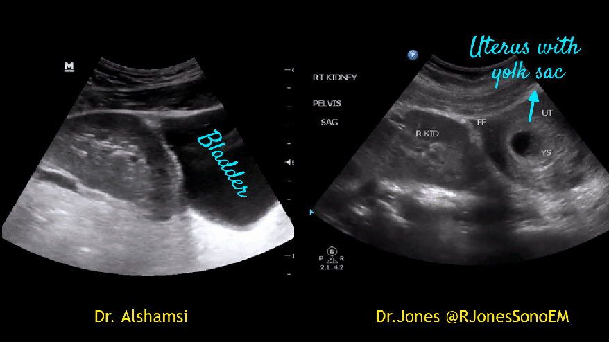
A middle-aged patient was seen in the nephrology clinic for evaluation of chronic kidney disease. No prior imaging was available. Patient was asymptomatic and there was no significant renal-related history except for occasional urinary tract infections. As a part of physical examination, medical resident tried to POCUS the kidneys but was able to find only the left one. It is a bit unusual because right kidney is generally easier to find as liver provider a better window. Of note, the resident did not scan the bladder. We rescanned the patient and still could not find the kidney in right upper quadrant. Next, scanned the midline to make sure we are not missing a horseshoe kidney. Still nothing. Then we moved to pelvic area and there it is! A right pelvic kidney was seen right next to the urinary bladder. It is pretty much like a transplanted kidney.


Pelvic kidney is an ectopic kidney, which essentially means that it did not ascend to the retroperitoneal renal fossa beyond the pelvic brim. In a significant proportion of patients, ectopic kidneys ae associated with vesicoureteral reflux other genitourinary abnormalities. Pelvic kidneys are usually smaller in size, have variable rotation, extrarenal calices and multiple sources of blood supply. Blood supply can be from a single artery arising at or just distal to the bifurcation of the aorta or dual (one from distal aorta, one from common iliac) or even triple (internal iliac in addition). Our patient possibly had dual supply though it is difficult to determine on a POCUS exam; see images below. Nevertheless, further imaging was not ordered as there were no symptoms and urinalysis was bland.


Here are two more examples. In females, a palpable pelvic kidney might be confused with a mass leading to unnecessary CT/MRI.