Infographics
Topics of general interest:


Focused echocardiography: general stuff


Cardiac views and chambers: anatomy


Cardiac views and chambers: Functional assessment


Cardiac chamber quantification


Pocket guide for pericardial tamponade


Diastolic dysfunction


cause the cardinal symptoms of heart failure. Parameters commonly measured to assess different aspects of LV filling pressures are indicated in brown.

Myocardial segments


Valves (swipe left or right)


Venous Doppler


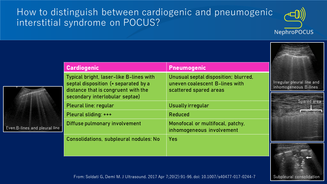
Lung ultrasound:


Abdomen and others:


