Horseshoe Kidney
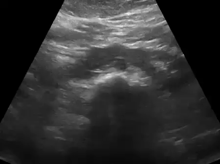
Horseshoe kidney is the most common renal fusion abnormality characterized by fusion of one pole of each kidney, typically the lower pole. The connection or the isthmus lies in the mid-line or sometimes slightly lateral to the mid-line and can be composed of renal parenchyma or just fibrous tissue. The renal collecting systems remain separate. Below anatomic and sonographic images demonstrate relations of the horseshoe kidney. IVC and aorta are important landmarks.


This anomaly is usually detected in childhood because of urinary tract infections, renal stones or hydronephrosis. However, in asymptomatic cases, it might be found incidentally during adulthood. When you don’t see lower poles of the kidneys clearly despite making sure there is no overlying bowel gas, scan in the mid-line. Sometimes, the isthmus of a horseshoe kidney can be interpreted as a mass arising from the kidney or the retroperitoneum if the mid-line scan is not performed.
Also note that the abnormal rotation of the kidneys shifts the renal pelvis and ureter anteriorly. Therefore, the location of hydronephrosis in these cases can cause confusion. Here are some images shared by Dr. Jones demonstrating hydronephrosis of the right moiety due to a distal ureteral stone (stone not shown here).


Here is a nice example showing stone in the right moiety of the horseshoe kidney shared by Dr. Zoran Paunic.


1 Comment »