Hiatal hernia on Focused Cardiac Ultrasound
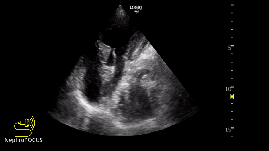
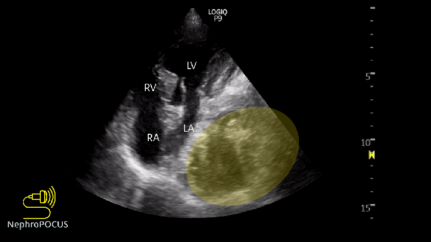
This apical 4-chamber view is obtained from a patient with shortness of breath being managed with escalation of diuretic therapy, which eventually led to hypovolemic hyponatremia. What does it show? What is the heterogeneous area adjacent to the left atrium?
It is hiatal hernia with gastric content. Patient has a long-standing history of gastroesophageal reflux and was told that their ‘stomach moved up into the chest’. LA compression by the hernia may cause dyspnea by increasing the pulmonary venous pressure leading to interstitial edema. In extreme cases, hemodynamic instability can result from impaired left ventricular filling. In addition, some patients may present with arrhythmias such as atrial fibrillation and heart block due to the pressure effect of hernia. Below are the labeled image and relevant illustrations.


The hernia is also seen on the right lateral scan while we are evaluating for pleural effusion or pulmonary edema (image below). It may be confused with atelectatic lung commonly seen in cases of pleural effusion. However, the structure seen here is much larger compared to the size of effusion, looks heterogeneous with air-fluid contents and appears to be lined by peritoneum. Remember, air is white, and fluid is black on ultrasound. On the other hand, atelectatic lung would look more like a liver with white dots inside (static air bronchograms).


Chest X-ray and CT scan are shown for better anatomic orientation. However, these were not from the exact same day as POCUS. CT shows gastric and transverse colon contents in the hernia.


Hiatal hernia is often well visualized on the parasternal long-axis echocardiographic view, where it may appear as a posterior structure adjacent to the left atrium. In our patient, however, obtaining an adequate parasternal window was challenging because of significant chest wall abnormality.
Below is an illustrative example from a different patient demonstrating a hiatal hernia on the parasternal view, along with corresponding CT imaging for correlation. At first glance, an inexperienced POCUS user might mistake this for a left pleural effusion, especially given the expected location discussed earlier. However, a closer look reveals several subtle clues suggesting otherwise. Unlike the relatively anechoic appearance of a typical pleural effusion, this structure appears more hazy and ill-defined. Part of that haziness results from dirty shadowing caused by the gas. In addition, the heterogeneous appearance does not resemble the classic image of a floating or atelectatic lung within pleural fluid. Instead, the mixed echogenicity arises from varying interfaces between solid material, fluid, and gas within the hernia.


Below are two nice images from the stated reference demonstrating achalasia on parasternal views, including the parasternal long-axis view and the parasternal short-axis view at the aortic valve level. Note the dilated esophagus located posterior to the LA. A hiatal hernia would appear very similar. After the patient consumed a carbonated beverage, repeat imaging demonstrated mobilization of the heterogeneous material within the dilated esophagus, with a noticeable increase in the anechoic (fluid-filled) component. In addition, compression of the LA appeared more pronounced following further esophageal distension.
This can serve as a useful bedside trick to help distinguish gastrointestinal structures and contents from other mediastinal pathologies such as tumors or even pleural effusions.


Excellent teaching and very high quality images
Thank you.
TREMENDO CASO PARA EL APRENDIZALE