IVC POCUS: one view is no view
We have previously discussed that longitudinal view of the IVC is subject to cylinder effect and it is a good idea to visualize it in short axis as well. However, it does not mean that its acceptable to examine just the short axis. In this example, short axis views of IVC obtained from the same patient show different degrees of collapsibility. There is no obvious change in respiratory effort of the patient (which can alter collapsibility). How would you grade right atrial pressure now? Intermediate (8 mmHg) or high (15 mmHg)?


Only way to know what’s going on is to look at the vessel in long axis.
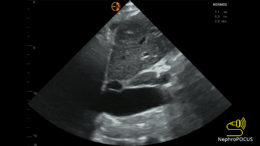
Long axis view shows vigorous diaphragmatic pull (false collapse), while in reality, IVC is minimally collapsing, which can be nicely appreciated without using M-mode. Dotted lines show approximate plane of the above transverse images. If you observe closely, Figure 1 shows more prominent hepatic veins. Its always better to visualize IVC in both planes and measure the diameter slightly inferior to hepatic vein-IVC junction.

Below is a different case discussing another pitfall of isolated transverse IVC POCUS