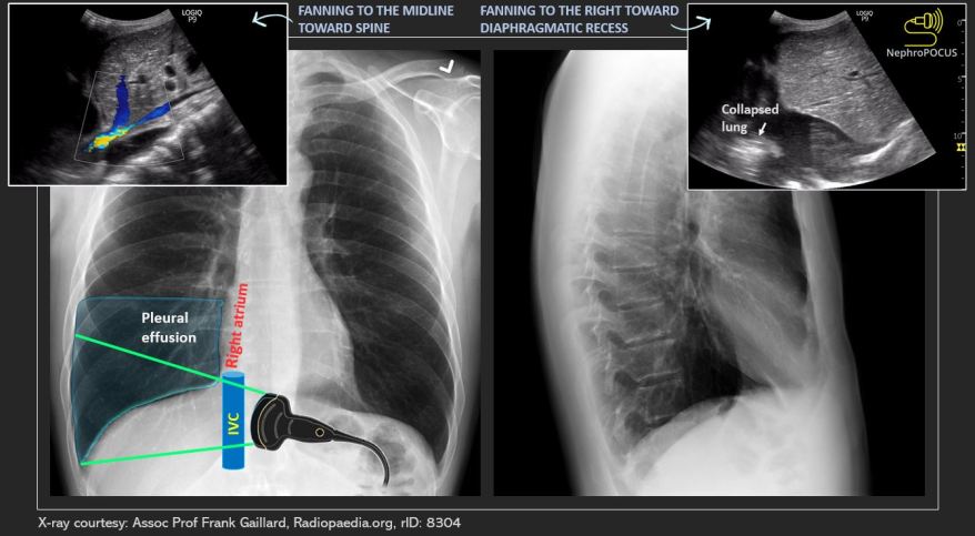
IVC and the right pleural effusion
In a twitter poll asking what the arrow was pointing to in this image, only ~54% answered correctly.


The correct answer is right pleural effusion. Below illustration explains why the pleural fluid appears ‘beneath’ the IVC in the angle we are scanning.


Two more examples:


As we’ve discussed before, pleural effusion, especially the right one is often visible from the subxiphoid window. Below image, captured with the probe angled superiorly in that window with orientation marker to the left, demonstrates a right pleural effusion, ascites, and a small pericardial effusion.


Below is another poll testing the same concept using a different image. This time also, many people got it wrong. Anatomic correlation below.


Below is another good example of a right pleural effusion visualized from multiple windows.


2 Comments »