Maternal Physiologic hydronephrosis in Pregnancy
Pregnancy-related hydronephrosis, more precisely physiologic maternal renal pelvis dilatation is common and the incidence is estimated to be as high as 80%. The dilatation of the pelvis and ureter typically develops toward the end of second trimester and disappears within a few weeks after delivery. It is more common in primigravid women and usually more prominent on the right side. Etiology includes extrinsic ureteral compression by the gravid uterus on top of reduced ureteral smooth muscle tone and peristalsis due to progesterone effect.
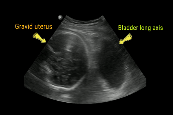
Right sided predominance may be due to dextrorotation of the uterus by the sigmoid colon, kinking of the ureter as it crosses the right iliac artery, and/or proximity to the right ovarian vein. Following figure gives an idea of the anatomy.

In these cases, it is often difficult to trace the ureter to the point of obstruction. However, in pathologic hydronephrosis, one may visualize the source of obstruction (e.g. stone). In addition, if the pelvic diameter exceeds 10 mm, pathologic dilatation should be suspected.
Here is a case of mild bilateral hydronephrosis in a primigravid pregnant woman during third trimester. Note weak left ureteral jet compared to normal. Color Doppler could not pick up the right jet despite maintaining the probe position for approximately 2 minutes consistent with obstruction.

Thanks. Good article and easy to understand