Venous valves
Encountering venous valves is not uncommon, and they can potentially be confused with pathology such as dissection by uninformed clinicians. In nephrology practice, internal jugular vein (IJV) valves are often observed when assessing right atrial pressure or placing a dialysis catheter. In fact, IJV valves are present in more than 90% of the population. These valves, located deep in the neck, are better visualized with a linear transducer with a smaller footprint (or a hockey stick probe) and in patients with a dilated IJV. Below are a couple of examples and an illustration demonstrating the position of IJV and subclavian valves.


Below is a nice M-mode image across the valve, illustrating that the valve remains open, allowing blood flow into the right atrium throughout most of the cardiac cycle, including both systole and diastole.

Being the sole valve between the brain and the heart, it plays a crucial role in preventing the backward flow of venous blood. Occasionally, valve damage during the insertion of a guidewire can create a nidus for thrombus formation. If you encounter ‘resistance’ while advancing the guidewire during catheter placement, consider the valve. In such cases, trying the ‘head-up’ position (instead of Trendelenburg) is recommended because the favorable gravitational gradient in an upright posture keeps the valve open throughout the cardiac cycle, making it easier to pass the guidewire. Below is an example illustrating the guidewire passing through the valve leaflets in the long-axis view. The short-axis view depicts needle entry through the valve as the operator follows the needle tip.


Veins in the lower extremities also contain valves, which can be encountered during deep vein thrombosis (DVT) assessment. These clips show a femoral vein valve in long and short axes. The adjacent Foley balloon-like structure in the short axis is a heavily calcified femoral artery, a common finding in patients with end-stage renal disease. In this case, the femoral vein is actually more pulsatile than the artery due to tricuspid regurgitation and pulsatile flow.

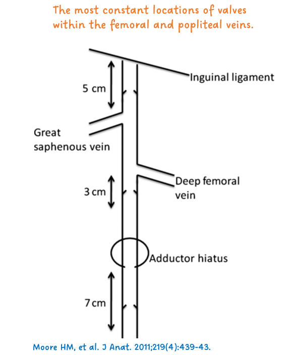
Shown below is a popliteal vein valve along with an illustration highlighting the typical locations of venous valves in the lower limb.