

In the previously posted videos and articles, we talked about the normal Doppler patterns of major abdominal veins used for VExUS grading and how they transition with worsening congestion. Here are some flashcards depicting the same.
Hepatic vein waveform. If you need a refresher on the wave genesis, watch this 1-minute slideshow.


Renal parenchymal vein


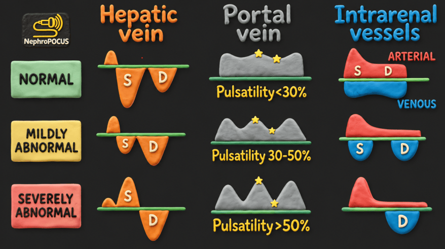


VExUS card with moving waveforms:

eVExUS or the extended VExUS

Below is a graphic illustrating congestion across various venous waveforms. It doesn’t include the actual scoring system, but gives a more visual representation of the changes, including the femoral vein. Green waveforms are normal, while orange waveforms are abnormal, with the arrows indicating progressively worsening congestion.

Below: Modified VExUS incorporating femoral vein Doppler (based on Joyal et al., PMID: 41931445). This approach essentially replaces intrarenal Doppler with the femoral vein stasis index and introduces a few additional modifications, including incorporation of IVC collapsibility and defining severe hepatic vein abnormality as either S-wave absence (i.e., S/D = 0) or S-wave reversal (i.e., S/D <0).

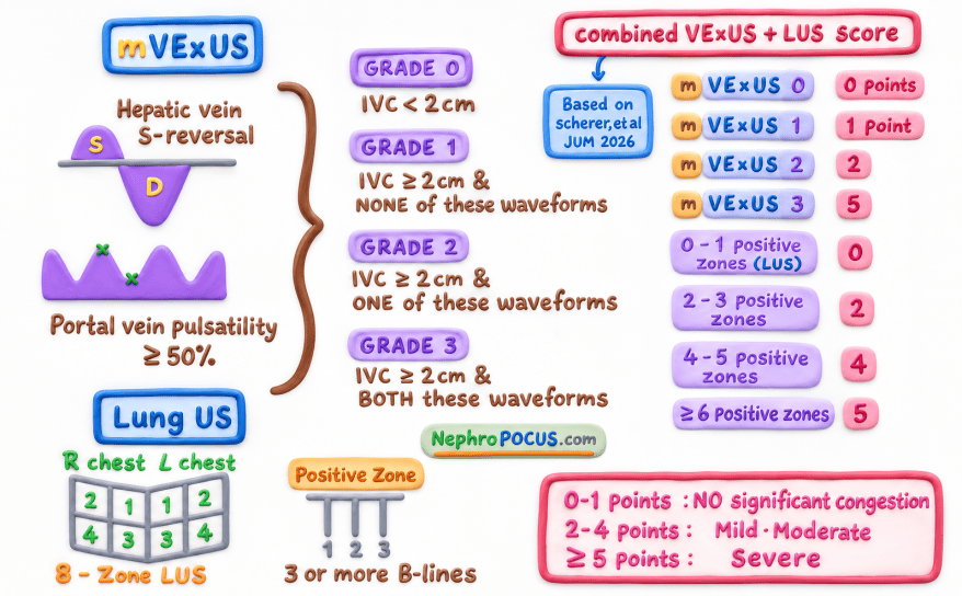
Below are two additional scoring systems:
- mVExUS (modified VExUS): Basically ‘simplified’ VExUS without the intrarenal venous Doppler.
- Combined mVExUS–LUS score: Combines mVExUS with lung ultrasound to capture both right- and left-sided congestion.

A gentle reminder:
